by Daniela Dominguez and Dr. Christopher Cirino
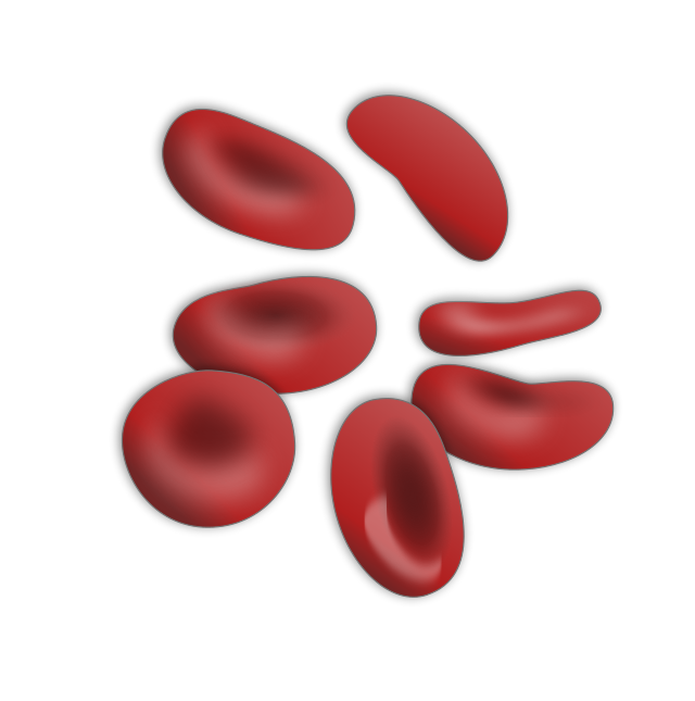
Red blood cells are the most abundant cells in the body. They transport oxygen from the lungs to every cell in our body. When the red blood cell count is low, it is referred to as anemia. The most common type of anemia is iron deficiency anemia. We all have heard about iron deficiency anemia, but what is it? And why is it important to determine its underlying cause?
Case: A 55-year-old man came to be seen by a primary care doctor for an initial visit with a complaint of fatigue. He did not report any obvious blood loss, that is, he did not mention any bloody stools or other causes. He had no weight loss or distress. The examination showed some spooning of his nails and mild pallor. A rectal exam showed a palpable firm nodule suggested. He was advised and given a referral for a colonoscopy. The bloodwork showed evidence of mild iron deficiency anemia with a hemoglobin/hematocrit count of 10 and 30 (normal > 14 and 42) and low Mean corpuscular volume (MCV).
He did not back to his primary care and was not seen by gastroenterology. Two years later, he came back to the office complaining of more severe fatigue, weight loss, and dark tarry stools. He now had marked pallor and a firm mass on his rectal exam. His blood count was low with a hemoglobin and hematocrit 7 and 23. He was admitted to the hospital and was found to have an advanced sigmoid colon cancer with partial obstruction on his CT scan.
What is iron deficiency anemia (IDA)?
Two major types of anemia exist based on the red blood cell size: macrocytic (large cells) and microcytic (small cells). With iron deficiency anemia, the drop in hemoglobin leads to small red blood cells. Iron deficiency is the most common cause of anemia worldwide. In the US, the prevalence of IDA is 2 percent of adult males, 9 percent of non-Hispanic white females, and 20 percent of black and Hispanic females (31). In blood tests, the mean corpuscular volume measures the average red blood cell size; in IDA, the MCV is less than 80fL (normal MCV 80-95fL).(1)

Why does the lack of hemoglobin make the red blood cells smaller?
Mature red blood cells, or erythrocytes, can circulate in the system for approximately 115 days (31). Red cells develop in the bone marrow from their precursors, erythroblasts. The larger erythroblasts divide and mature into red blood cells, which lose their nuclei and are ready to leave the bone marrow. Red cell development and turnover become dysfunctional in the setting of deficient iron stores, resulting in pale and small red cells.
What causes IDA?
Iron deficiency anemia occurs in any situation that leads to low iron levels in the body: decreased iron intake or absorption, iron loss, and increased iron demand. (4) IDA is the most common nutritional deficiency worldwide. (5)(6).
Iron is an essential element for the survival of all cells. The body monitors iron stores closely. Aside from the usual iron loss from skin and mucosal cell turnover, blood loss can accelerate the depletion of stores.
In the small intestines, the duodenum and initial part of the jejunum absorb iron; in normal conditions, only 10% of the consumed iron enters. During pregnancy and other depletions states, the amount absorbed can increase 3 to 5 times.
Iron in our diet comes in two forms: heme and nonheme iron. Heme iron comes from meat, and its absorption does not depend on other dietary factors. Nonheme iron comes from plant-based ingredients, and to be absorbed requires the stomach’s acidity. The co-consumption of certain foods such as fiber, calcium, tea, coffee, or wine can inhibit iron. (7)
Men and non-menstruating women lose 1mg of iron daily. Women of reproductive age lose around 0.6 to 2.5 mg of iron per day, and a 60kg woman loses approximately 10mg of iron each menstruation cycle, but in menstruation problems, a woman can lose more than 42mg of iron per cycle. (4)
Since iron deficiency anemia occurs in more severe conditions, all patients should receive a thorough investigation of the potential sources. The table below lists some of the causes of IDA. A known and relatively common source of bleeding, e.g., hemorrhoids, does not always mean the work-up is over, particular in someone with more severe anemia.
Causes of Iron Deficiency Anemia
| Iron Loss | Known Blood Loss | Abnormal uterine bleeding, gastrointestinal bleeding (long-term use of aspirin or other nonsteroidal anti-inflammatory drugs, peptic ulcer disease, angiodysplasia, gastric antral vascular ectasia, Cameron ulcer), blood donation, hematuria, epistaxis |
| Iron Loss | Occult Blood Loss | Colon, gastric, and esophageal carcinoma, small bowel tumors, ampullary carcinoma, hematuria, metastatic cancer |
| Iron Absorption Issues | Celiac disease, gastrectomy, helicobacter pylori infection, bacterial overgrowth, intestinal resection. |

What are the signs and symptoms of IDA?
Fatigue is the most common symptom. (Here is an article on work-up of fatigue) Other signs and symptoms may occur, like shortness of breath, pallor, headache, and lightheadedness. (9)(10)(3) Heart issues like angina manifest due to the increased demand for oxygen from the heart, especially in patients with preexisting cardiovascular disease. (1)
However, most patients with iron deficiency anemia have no apparent signs and symptoms if it is mild or moderate.
Pale conjunctiva and skin can suggest anemia; however, the lack of pallor does not rule out anemia. (11) Other signs and symptoms like spoon nails (koilonychia), swollen tongue (glossitis), or trouble swallowing (dysphagia) are not common in developed countries. (12) Another symptom of anemia is pica, which refers to the compulsive eating of nonedible substances (like dirt or ice). It presents more often in pre-adolescents and pregnant women. Iron therapy usually cures the behavior. (13)
Iron deficiency anemia plays a role in the pathophysiology of restless leg syndrome. The incidence of iron deficiency anemia increases 8 to 10-fold among women with restless leg syndrome. Screening for IDA is crucial in these patients (14)
Most patients with mild to moderate iron deficiency anemia have no apparent signs and symptoms
How to diagnose iron deficiency anemia?
Diagnosis starts with a Complete Blood Count (CBC) with a hemoglobin level two standard deviations below normal for age and sex. (15) The CBC will show small cells (Low MCV and Low Mean Corpuscular Hemoglobin (MCH). The CBC will also show an increased value in the Reb Blood Cell Distribution Width (RDW), reflecting high red blood cell size variation due to poor erythropoiesis.
Iron deficiency anemia will also have a low reticulocyte count. Reticulocytes are immature red blood cells in the bloodstream and come from the bone marrow, and in two days become fully mature. Since the making of red blood cells (erythropoiesis) is impaired, reticulocyte count will decrease due to the lack of iron. (8)(2)
Up to 40% of people with iron deficiency anemia have a normal MCV. One should still consider iron deficiency anemia. Normocytic anemia precedes the development of microcytic anemia in IDA, as iron stores fall. (6)(16)
The next step to clarifying the type of anemia is to measure blood iron levels with an iron saturation level. The ferritin level shows iron stores. It accurately estimates iron deficiency (less than 30ng per mL). Ferritin levels greater than this are usually not IDA; more than 100 ng per mL generally exclude iron deficiency anemia. (17–20) Ferritin is also an inflammatory marker and can sometimes be elevated in the setting of inflammation, confounding interpretation.
Additional tests are helpful in iron deficiency anemia: the Total Iron Binding Capacity (TIBC) increases because the low iron stores increase empty receptors. A high TIBC suggests an iron deficiency in the setting of low iron saturation and ferritin levels.
The peripheral blood smear shows microcytic, hypochromic red blood cells with anisocytosis (RBC unequal in size) and poikilocytosis (RBC unequal in shape).(8)
| Lab Results of Iron Deficiency Anemia |
| Microcytic Anemia (Low MCV) Low Ferritin Level Low Iron Saturation High TIBC |
Who should be screened for iron deficiency anemia?
Men and postmenopausal women do not need regular screening. Doctors screen any individual with signs and symptoms of fatigue, dizziness, and restless legs or concerning physical findings of pallor, spooning nails, or positive occult blood in the stool. Diagnostic tests should be performed only with signs and symptoms or incidental laboratory tests that suggest anemia. (25)
Screening for iron deficiency anemia is recommended in all pregnant women according to The American Academy of Family Physicians, U.S. Preventive Services Task Force, Centers for Disease Control and Prevention, and The American College of Obstetricians and Gynecologists, the last also recommend iron deficiency supplementation if iron deficiency is confirmed. In pregnancy, anemia is diagnosed with hemoglobin levels of less than 11g per dL in the first trimester or less than 10.5g per dL in the second trimester. A maternal hemoglobin level of less than 6g per dL can be fatal to the fetus. (15)(21)(22)(23)
Pediatricians screen children at one year of age and evaluate them if they have risk factors (low birth weight, prematurity, exposure to lead, exclusive breastfeeding beyond four months of life, and weaning to whole milk and complementary foods without iron-fortified foods) (24)
What is the Treatment or IDA
It is essential to address an underlying condition causing iron deficiency anemia since it can be serious (e.g., color or uterine cancer). Primary care doctors refer individuals with IDA to a gynecologist or gastroenterologist for a thorough evaluation and treatment.
Oral iron therapy
Iron sulfate (325mg = 65mg elemental iron) is most commonly used and is taken once or twice daily. Follow-up blood tests can check progress after 1 month. Therapy should continue for three months after the anemia corrects, providing that the cause is established (see Table above).
Oral iron therapy can cause various side effects, such as epigastric discomfort, diarrhea, nausea, constipation, and black stools, making it hard for patients to adhere to treatment. Symptoms may improve if taken with meals, but the absorption decreases. The use of proton pump inhibitors reduced the absorption as well as having factors that reduce gastric acid: chronic atrophic gastritis, recent gastrectomy, or vagotomy.(5)(16)(24)(26,27)
IV Iron Therapy
Doctors reserve IV therapy for patients who cannot tolerate oral therapy or have serious gastrointestinal effects, worsening symptoms of inflammatory bowel disease, unresolved bleeding, renal failure-induced anemia treated with erythropoietin, or insufficient oral absorption (e.g., celiac disease).
Iron sucrose and sodium ferric gluconate are preferred due to greater bioavailability and lower anaphylaxis incidence than iron dextran. Side effects include headache, nausea, and diarrhea. Ferumoxytol supplies 510mg of elemental iron per infusion, allowing more iron in fewer infusions than iron sucrose.(6)(28)(29,30)
Take Home Points
Iron deficiency anemia is the result of depleted iron stores, usually from decreased iron intake or blood loss. Hemoglobin is underproduced, leading to dysfunctional production of red cells in the bone marrow. It manifests as fatigue, headache, pallor, and restless leg syndrome. Identification of iron deficiency anemia requires a careful assessment as to its underlying cause. This may include further testing, such as colonoscopy and imaging studies like a computed tomography (CT) or pelvic ultrasound.
Bibliography
1. BARONE, J. AND CASTRO MA. USMLE® step 1 lecture notes 2016: Pathology. New York: Kaplan-Medical; 2016.
2. ROBBINS, S. L., ASTER, J. C., PERKINS, J. A., ABBAS, A. K. AND KUMAR V. Robbins basic pathology. 10th ed. Philadelphia: Elsevier; 2018.
3. Sattar HA. Fundamentals of Pathology. 2018 Editi. Chicago: Pathoma LLC; 2018.
4. Rodgers JPGDAABEGAFLRMMGM. Wintrobe’s Clinical Hematology. 14th editi. Wolters Kluwer Health;
5. World Health Organization. Iron deficiency anaemia: assessment, prevention and control [Internet]. 2001. Available from: https://www.who.int/publications/m/item/iron-children-6to23–archived-iron-deficiency-anaemia-assessment-prevention-and-control
6. Johnson-Wimbley TD, Graham DY. Diagnosis and management of iron deficiency anemia in the 21st century. Therap Adv Gastroenterol. 2011 May;4(3):177–84.
7. Uzel C, Conrad ME. Absorption of heme iron. Semin Hematol. 1998 Jan;35(1):27–34.
8. Tao, Le; Vikas, Brushan; Maniver, Deol; Gabriel R. First Aid for the USMLE Step 2CK. 10th ed. The McGraw-Hill Education; 2019.
9. Powers JM, Buchanan GR. Disorders of Iron Metabolism: New Diagnostic and Treatment Approaches to Iron Deficiency. Hematol Oncol Clin North Am [Internet]. 2019;33(3):393–408. Available from: https://www.sciencedirect.com/science/article/pii/S088985881930022X
10. Elnicki DM, Shockcor WT, Brick JE, Beynon D. Evaluating the complaint of fatigue in primary care: diagnoses and outcomes. Am J Med. 1992 Sep;93(3):303–6.
11. Sheth TN, Choudhry NK, Bowes M, Detsky AS. The relation of conjunctival pallor to the presence of anemia. J Gen Intern Med. 1997 Feb;12(2):102–6.
12. Cook JD. Diagnosis and management of iron-deficiency anaemia. Best Pract Res Clin Haematol. 2005 Jun;18(2):319–32.
13. Borgna-Pignatti C, Zanella S. Pica as a manifestation of iron deficiency. Expert Rev Hematol. 2016 Nov;9(11):1075–80.
14. Kolukisa M, Soysal P, Gületkin TÖ, Karatoprak C, Bilgen HR, Gürsoy AE. Restless Leg Syndrome/Willis-Ekbom disease in women with iron deficiency anemia. Ideggyogy Sz. 2016 Sep;69(9–10):356–60.
15. Siu AL. Screening for Iron Deficiency Anemia and Iron Supplementation in Pregnant Women to Improve Maternal Health and Birth Outcomes: U.S. Preventive Services Task Force Recommendation Statement. Ann Intern Med. 2015 Oct;163(7):529–36.
16. Ioannou GN, Spector J, Scott K, Rockey DC. Prospective evaluation of a clinical guideline for the diagnosis and management of iron deficiency anemia. Am J Med. 2002 Sep;113(4):281–7.
17. Mast AE, Blinder MA, Gronowski AM, Chumley C, Scott MG. Clinical utility of the soluble transferrin receptor and comparison with serum ferritin in several populations. Clin Chem. 1998 Jan;44(1):45–51.
18. Knovich MA, Storey JA, Coffman LG, Torti S V, Torti FM. Ferritin for the clinician. Blood Rev. 2009 May;23(3):95–104.
19. Galloway MJ, Smellie WSA. Investigating iron status in microcytic anaemia. BMJ. 2006 Oct;333(7572):791–3.
20. Skikne BS, Punnonen K, Caldron PH, Bennett MT, Rehu M, Gasior GH, et al. Improved differential diagnosis of anemia of chronic disease and iron deficiency anemia: a prospective multicenter evaluation of soluble transferrin receptor and the sTfR/log ferritin index. Am J Hematol. 2011 Nov;86(11):923–7.
21. World Health Organization. Assessing the iron status of populations: including literature reviews: report of a Joint World Health Organization/Centers for Disease Control and Prevention Technical Consultation on the Assessment of Iron Status at the Population Level, Geneva, Switzer. 2004;
22. Recommendations to prevent and control iron deficiency in the United States. Centers for Disease Control and Prevention. MMWR Recomm reports Morb Mortal Wkly report Recomm reports. 1998 Apr;47(RR-3):1–29.
23. ACOG Practice Bulletin No. 95: anemia in pregnancy. Obstet Gynecol. 2008 Jul;112(1):201–7.
24. Baker RD, Greer FR. Diagnosis and prevention of iron deficiency and iron-deficiency anemia in infants and young children (0-3 years of age). Pediatrics. 2010 Nov;126(5):1040–50.
25. Bermejo F, García-López S. A guide to diagnosis of iron deficiency and iron deficiency anemia in digestive diseases. World J Gastroenterol. 2009 Oct;15(37):4638–43.
26. Ioannou GN, Rockey DC, Bryson CL, Weiss NS. Iron deficiency and gastrointestinal malignancy: a population-based cohort study. Am J Med. 2002 Sep;113(4):276–80.
27. Ajmera A V, Shastri GS, Gajera MJ, Judge TA. Suboptimal response to ferrous sulfate in iron-deficient patients taking omeprazole. Am J Ther. 2012 May;19(3):185–9.
28. Goddard AF, James MW, McIntyre AS, Scott BB. Guidelines for the management of iron deficiency anaemia. Gut. 2011 Oct;60(10):1309–16.
29. Maslovsky I. Intravenous iron in a primary-care clinic. Am J Hematol. 2005 Apr;78(4):261–4.
30. Silverstein SB, Rodgers GM. Parenteral iron therapy options. Am J Hematol. 2004 May;76(1):74–8.
31. Franco M. Measurement of Red Cell Lifespan and Aging. Transfus Med Hemother. 2012; 39: 302-307.
32. Killip S, et. al. Iron Deficiency Anemia. American Family Physician. 2007; 75(5): 671-678.


Leave a Reply