History
51 yo with increasing shortness of breath, cough and weakness over a period of a few weeks.
- No previous significant past medical history
- Low grade fever
- Works as a custodian and was subjected to a lot of second-hand smoke
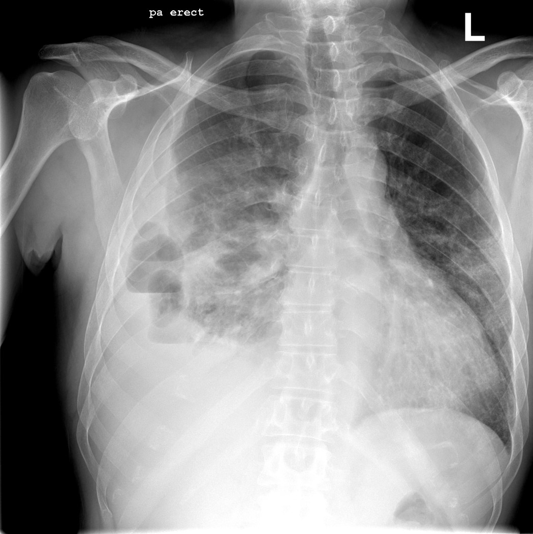
His initial CXR:

Hospital Course
- Initially placed on Rocephin, Levaquin and Doxycycline
- Started on Solumedrol IV for several days.
- Initial culture of sputum: Normal flora; Influenza culture: Negative
- Had a bronchoscopy 5 days after admission for worsening respiratory distress: negative stains for Pneumocystis (PJP). Gram stain showed “few Gram positive cocci. Culture grew “yeast and normal respiratory flora”


On account of CXR findings, a CT scan was ordered:


Question 1: What process is likely being depicted in this CT scan?
- The patient underwent a Video-Assisted Thoracoscopic surgery (VATS) with decortication on account of the findings)
- Cardiothoracic (CT) surgery found an area of necrotic tissue in the setting of the Right lower lobe process and a thick pleural peel.
Pathology contacted the physician after their review:


Question 2: What is the name of the specialized stain shown above? Why is it used?
Question 3: What is shown using this specialized stain?
Question 4: What do you suspect was the diagnosis and likely risk factors? Hint: Risk factors are from history, hospital course and discovered (at least 3).
Click for the answer to this week’s ID Case Report Challenge.


Leave a Reply