
Answer: Mycobacterium tuberculous colitis
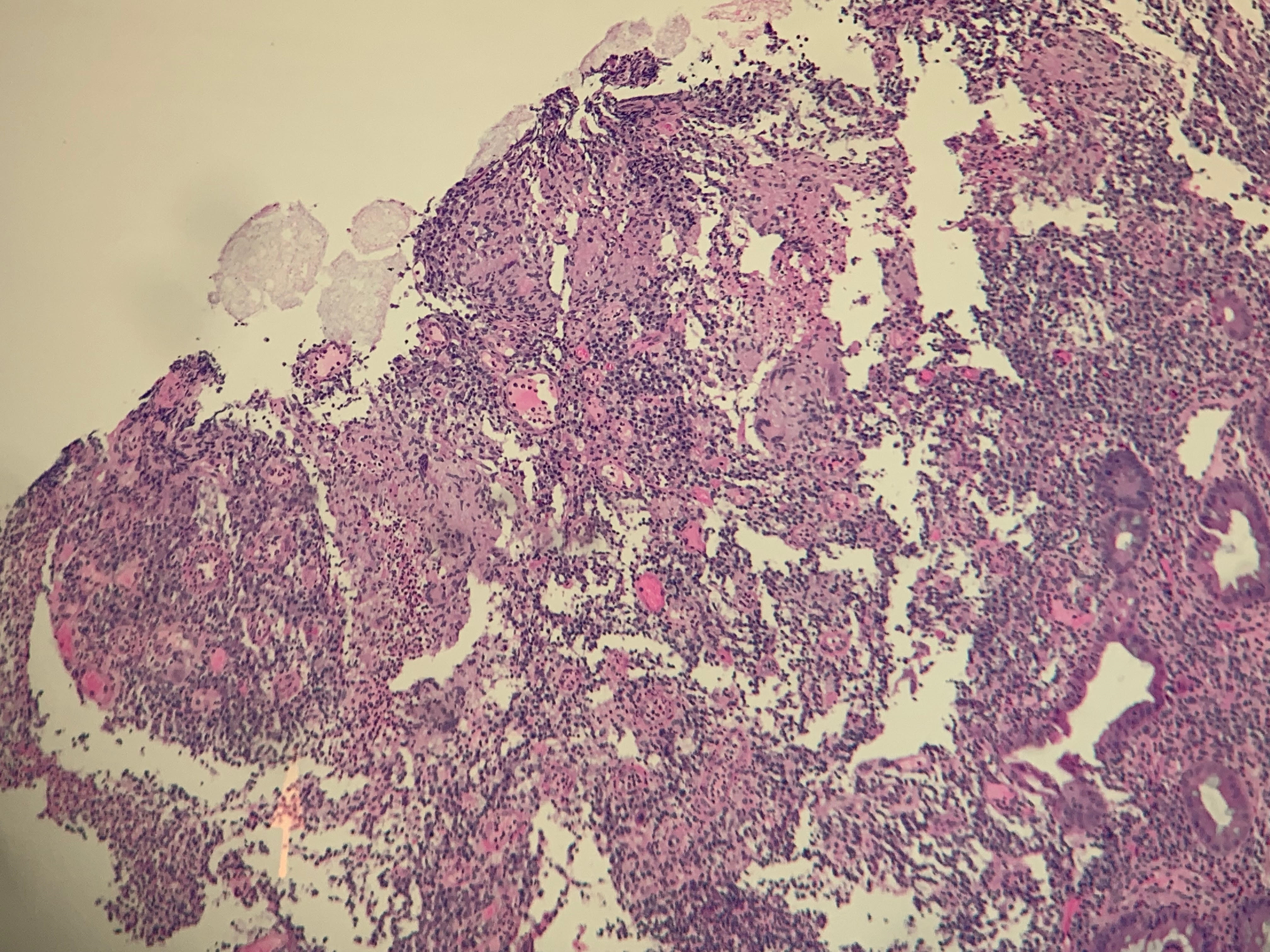
- What structures are visualized on the H & E stains? The structures are subtle but relate to non-caseating granulomas found in the colonic mucosa
- What is the special stain that was used and what is shown? The stain is known as a FITE stain which stains the bacterial cell wall of Mycobacteria species, which have cell walls containing mycolic acids – lipid-rich. The organism stains pink/red and cellular material stains blue.
- What additional studies are recommended to pursue?
- Quantiferon test: POSITIVE
- Chest Radiograph: NEGATIVE
- Efforts were made to get a specimen for culture and identify the organism as Mycobacterium tuberculosis
- Colonic mucosa pathology – requested to be sent for Mycobacteria tuberculosis PCR – NEGATIVE
- Stool for Acid fast bacilli stain x3 – these were NEGATIVE
- Repeat colonoscopy for fresh samples — this was performed – however, no fresh specimen was submitted.
- Sputum for AFB and culture and MTB-PCR: Smears x 3 were NEGATIVE but PCR was POSITIVE
- What is the working diagnosis in this patient? The working diagnosis is tuberculous colitis with presumed pulmonary site. The smears were negative x 3 but the Mycobacterium tuberculosis PCR test was positive. Cultures were set up and the patient was begin on four drug regimen with isoniazid, rifampin, pyrazinamide and ethambutol.
Tuberculous colitis
The gastrointestinal manifestations of active tuberculosis can be subtle to severe, with disease presenting at any level. It is likely that this process is more common in tuberculosis than discovered, found from 60-90% of post-mortem examinations of those with active tuberculosis. Likely, this is a result of the natural passage of bacilli through the gastrointestinal system from swallowed pulmonary secretions.
The colonic lesions may be seen as small, superficial erosions or pseudopolyps to a more severe ulcerative disease, even mimicking Crohn’s disease or ischemic colitis. The classic pathological finding of tuberculosis is caseating-necrosis, caseus Latin for “cheese”, and is only seen in about 50% of cases. It is also a finding seen in inflammatory bowel disease like Crohn’s disease. The difference between caseating and non-caseating is shown below and relates to presence or absence of cellular debris at the necrotic center.
Other than a work-up for military, or disseminated tuberculosis, treatment is similar to treatment of any active tuberculosis for approximately 6 or 9 months. Four active medications are used for two months in the induction phase as outlined above. Following this phase, cultures are usually available to determine drug sensitivity and, providing no resistance is detected, the regimen becomes isoniazid plus rifampin for the remainder of the therapy.


Leave a Reply